
|
PT Classroom - Imaging of Joint Injuries in Athletes ׀ by Cary Pallin, MD |

Dr. Cary Pallin attended the University of Arkansas where she received her medical degree and completed her residency in diagnostic radiology. In 2002, Cary and her husband, John Pallin, MD, who is also a Board Certified radiologist, started Kenosha Radiology Center (KRC). KRC is a comprehensive outpatient imaging center which utilizes the most up to date medical imaging technology. |

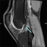
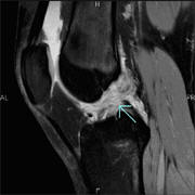
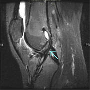
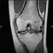
Medical imaging has become increasingly important in the diagnosis of joint injuries in athletes, providing prompt accurate diagnosis. X rays are often performed first to assess for fracture. In those without fracture a MRI is usually the next step. A MRI provides detailed images of the non-bony structures of the joint which are often injured, including ligaments, tendons, menisci, and cartilage.
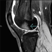
In an athlete with a knee injury, MRI can assess the status of the ACL (anterior cruciate ligament), the menisci, the medial and lateral collateral ligaments, the articular cartilage, and the patellar mechanism. It is also sensitive for occult fracture and bone contusion. These findings assist the orthopedic surgeon in determining the best course of treatment whether conservative or surgical and in preoperative planning.
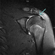
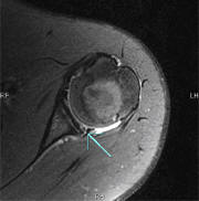
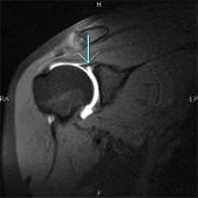
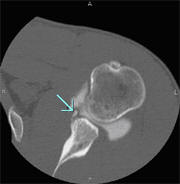
In the shoulder, MRI provides the best non-surgical evaluation of the important non-bony structures of the joint, including the rotator cuff tendons, labrum, and biceps tendon. Detailed evaluation of abnormalities of the labrum is enhanced by intra-articular injection of MR and/or radiographic contrast, an exam known as an arthrogram. This is a simple procedure that involves placement of a needle into the joint using fluoroscopic guidance and injection of the contrast agent, followed by post arthrogram imaging with MRI and/or CT. Post arthrogram CT images provide very high-resolution images of small structures.
MRI is similarly useful in evaluating wrist, elbow, hip, and foot and ankle injuries.
Displayed below is a comparison chart of some of the medical imaging modalities which are available and can assist the healthcare professional in determining a more accurate diagnosis for their patients. |
|||||||||||||||||||||||||||||||||||||||||||||||||||||||||
|
|
|
|
|
|
|
|


Terms & Conditions

