Legg-Calve-Perthes Disease is a pediatric disease which affects the femoral
head. The disease is an avascular necrosis of the femoral head always including
the capital femoris epiphysis. Legg-Calve-Perthes Disease (LCPD) affects 1 in
1200 children with boys more than girls, 4:1 (6). LCPD shows up mostly in the
Caucasian race. Most often the disease is unilateral but a small percentage of
children with LCPD do develop the disease in bilateral hips. Most children are
diagnosed between 6 to 9 years of age with a range from 2 to 12 years old (6).
The disease will manifest itself and recover within a year or a few. The disease
is also called coxa plan or osteochondritis of the femoral head.
The initial pathology which causes the avascular necrosis of LCPD remains
unknown. Doctors have hypothesized LCPD may be initiated by trauma, synovial
effusion, infection, vascular occlusion, constitutional predisposition, and
genetic factors (8). The cause may be described as a vascular tamponade of the
artery which supplies the femur. The femoral head and neck have limited blood
supply so it is more susceptible to ischemia. The first signs and symptoms of
LCPD may manifest at anytime in childhood. Some symptoms may be a limp or pain
in the hip or referred pain to the inner thigh and knee area. Some signs may
include limited hip range of motion especially limited hip abduction (7). There
is usually not a precipitating incident which causes the pain. The onset is
insidious in nature. Diagnosis of the disease must be done by radiographs, MRI,
or bone scan (6).
There are four stages where Legg-Calve-Perthes Disease progresses through. The
first stage lasts 6 to 12 months and the femoral head becomes more dense with
possible fracture of supporting bone (exact). Phase two consists of
fragmentation and reabsorbtion of the bone and takes one year or more. Phase
three is reossification when new bone has regrown and may take years. Phase four
is healing and may also take years (6).
The disease can affect part or all of the femoral head depending on severity of
the case. The prognosis may be better in younger children because the regrowth
of the femoral head is shaped better for congruency (6). The Catteral
Classification has four different groups to classify defined by radiograph
appearance in the greatest period of bone loss. An article by Van Dam et al.
concluded that the Catteral Classification is valid, reliable and simple to use
but needs to be classified after fragmentation of the capital epiphysis (9). The
Salter-Thomson Classification takes the Catteral Classification and makes it
only two groups for simplification purposes. Group A (formerly Catteral 1 and 2)
which is less than 50% of the femoral head is involved. Group B (formerly
Catteral 3 and 4) which is more than 50% of the femoral head is involved (6). A
better prognosis is Group A over Group B. The Herring Classification looks at
the lateral pillar of the ball of the femoral-acetabular ball and socket joint.
In group A, there is slightly density change and no loss of height in the
lateral 1/3 of the femoral head. Group B there is loss of height of less than
50% total height and lucency of the bone. Group C is when there is more than 50%
of lateral height (6).
As children grow there are many different types of diagnoses besides LCPD which
may cause hip pain in children. It is important to do a thorough history taking
and physical examination to help determine what the cause of the hip pain is to
differentially diagnose the disease. One of the first causes may be from a
septic hip joint in which rapid diagnosis is important so as not to cause
impaired blood flow to the joint. Major signs and symptoms of the sepsis will be
fever, pain, and inability to stand. Radiographic findings include widening of
the joint space. Treatment includes surgical drainage and antibiotics. Another
reason for pediatric hip pain may be osteomyelitis. Osteomyelitis is
inflammation of the bone and its structures secondary to infection. This
diagnosis may have a history of trauma. This is characterized by fluid in the
joint and signs include pain with standing, pain with hip range of motion, and
maybe a low grade fever. Treatment is rest and analgesics for pain which may
last for a few days. Another cause of hip pain may be slipped capital femoral
epiphysis which is a fracture of the growth plate leading to slipping of the
epiphysis. For 10-15 year olds, incidences in males are greater than in females.
The child may often be overweight, ambulate with foot externally rotated, and
have pain with internal rotation. Treatment is surgery to repair the joint.
Another cause of pediatric hip pain is Osteoid Osteoma which is a benign bone
tumor common in the demur and tibia. Treatment is surgery. Malignancy,
rheumatologic disease. And trauma are other causes of hip pain (2).
Treatment of Legg-Calve-Perthes Disease will differ depending on progression and
severity of disease. For mild LCPD treatment may only include physical therapy
exercises to maintain range of motion. Non-surgical treatment can include
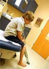
crutches for non-weight bearing for decreased pain. Braces and casts can help to
regain mobility of the hip joint. Braces may often help in hip abduction for
better joint congruency. Surgical treatments may include tenotomies or
osteotomies. Tenotomies may be needed to lengthen a shortened muscle due to
antalgic gait (6). Osteotomies are done to reposition the femoral-acetabular
joint for maximal congruency. Osteoomies are either done of the pelvis to
reposition a shelf superior to the joint or to the femur to reposition the joint
for congruency. The destruction of the bone from LCPD changes the shape of the
femoral head. A prospective study by Herring et al. looks at the effects of
treatment of Legg-Calve-Perthes Disease (3). The study looked at five different
treatments of LCPD which included no treatment, brace treatment, range of motion
exercises, femoral osteotomy, and innominate osteotomy. The study found that
lateral pillar classification and age of onset were determining factors in
outcome of the disease. There were no differences within conservative treatments
of no treatment, bracing, or range of motion. There were also no differences in
which osteotomy was performed. The study showed that treatment did not have a
great effect if the children were less than 8 at age of onset. If the age of
onset was greater than 8 years of age the treatments had a better outcome. In
those patients in Herring lateral pillar B classification, operative treatments
had better outcomes than non-operative treatment. In Herring C classification
the treatment outcomes were poor for operative and non-operative treatments (3).
One study by Westhoff et al. looked at the gait abnormalities that can happen
with LCPD. The study found a trendelenburg gait pattern or a compensated
trendelenburg gait pattern in children with LCPD. The trendelenburg gait pattern
consists of a drop in the contralateral pelvis in stance of the affected hip.
Compensated trendelenburg gait pattern results in an ipsilateral trunk lean to
the affected side. The compensated trendelenburg leads to a decreased hip
abductor moment arm which is correlated with a decrease in loading of the
ipsilateral hip joint (1). LCPD patients have weak hip abductors which lead to
the deviations in gait.
One article by Koob et al. looks at the growth of bone and cartilage following
ischemic osteonecrosis of the femoral head (5). The study produced ischemia in
the femoral head of piglets and looked at an 8 week follow-up and studied
different components of the bone. The bone core strength remained lower
throughout the eight week study and became progressively lower in the ischemic
head (5). The control for the study was the contralateral hip.
Patients with Legg-Calve-Perthes Disease may receive a variety of treatments
from conservative to surgical. Since disease is insidious in onset and
progressive, it may make any potential benefits from physical therapy hard to
determine. In the study by Herring et al. (3), they found no significant
evidence to support that physical therapy would help versus no treatment.
However, children with LCPD may have functional deficits such as an antalgic
gait with subsequent weakness and loss of range of motion. Physical therapy may
help maintain range of motion and hip strengthening which are two factors that
are decreased with this disease and worsen the antalgic gait. Patients will also
need gait training with their orthoses or crutches for proper use. They may also
benefit from aquatic therapy to allow for ambulation and strengthening exercises
with decreased body weight that they need to support due to the buoyancy effects
of the water. Physical therapy may also be beneficial after the disease has
taken course and the bone rebuilding and regrowth has taken place. The children
with LCPD will have disuse atrophy and will need to relearn gait so they no
longer limp. Any additional exercises and stretching may be added to bone
loading exercise to help the child back to pre-disease sate. Although physical
therapy may not be needed consistently throughout the course of the disease, it
may be beneficial at times for maintaining and regaining function.
Last revised: March 15, 2011
by Kristin Konicek, DPT


