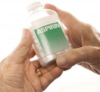
Acetylsalicylic
acid (ASA), commonly called aspirin, is a drug often used for the
relief of pain and inflammation. It is a relatively inexpensive drug
and is one of the most widely used peripherally acting analgesics in
the world (1). In addition, ASA is often one of the major oral
analgesics of choice for osteoarthritis (OA), a disease that is
characterized by articular cartilage deterioration (1). In some
other cases, however, additional research has indicated ASA might
inhibit the synthesis materials required for the maintenance and
repair of articular cartilage (AC). Although further research is
required to warrant these findings one would now have to question
the administration of ASA to individuals who have AC damage. The
purpose of this review is to recognize and analyze the research that
discusses the effects ASA has on AC.
Literature Review
Preliminary research in the past seems to support the use of ASA in
the inhibition of cartilage deterioration. A study conducted by
Simmons et al (2) showed that sodium salicylate (SS) was able to
prevent a decrease in the viscosity of chondromucoprotein (CMP), a
major macromolecular component of cartilage matrix. In their in
vitro study 13 pairs of extracts of human AC were taken from three
normal and nine patients with OA during surgery. These extracts were
than homogenized in normal saline at 4 degrees C and centrifuged.
After this process one half of each pair had SS added to them and
all samples were than incubated with 1% CMP at pH 7 for 3 hours with
frequent viscosity determinations. Results showed that the relative
viscosity reduction of CMP was nearly always less in those mixtures
containing SS (p<0.001). These are important findings, because in OA
catabolic enzymes are often released that breakdown CMP which is
needed for cartilage maintenance and growth (2). In interpreting
these results one should realize that the CMP used was prepared from
calf nasal cartilage which might not be similar enough to human CMP.
In addition, it is left unclear from the article’s methods where the
thirteenth pair of AC was obtained which makes one question if the
authors were detailed enough in describing their study.
Similar results on the protective effects of ASA against
degeneration of human AC was also found in a study conducted by
Christman et al. (3). In this study the researchers hypothesized
that salicylate can inhibit degradative enzymes in cartilage and
allow for synthesis to catch up with degeneration. For this study
they utilized individuals with recurrent lateral dislocations of the
patella who are receiving surgical correction after the second or
third occurrence. After the dislocation and 6-8 weeks before surgery
the individuals were randomly divided into two groups with the
individuals in one group instructed to take 3g of Bayer’s aspirin a
day, everyday, before the surgery, while the individuals in the
other group received no such instruction. During the surgical
procedures the researchers were allowed to inspect the cartilage
surface of the patella and rate it by the grading system developed
by Bentley, which was not described in the article. The results of
the study showed that of the 23 knees in the control group, only two
showed no evidence for chondromalacia. Strongly contrasting with
this, of the aspirin-treated group of 16 knees, 13 knees showed no
signs of chondromalacia. Although the authors obtained significant
statistical results, they did caution that a strict double blind
study should have been implemented to increase reliability and
validity. Other factors that the authors failed to mention that
could have also affected the reliability and validity of the study
was the adherence of taking the aspirin by the experimental group
and the control of extraneous variables for both groups such as the
amount of nutrition or rest each individual received. The number and
severity of dislocations for each person could have affected the
results as well.
A study by Roach et al (4) further supports the previously mentioned
studies on the positive effects ASA has on the degradation of
cartilage. In this study the authors compared the effects of
steroid, ASA and SS on AC. Rabbit knee joints were compressed to
produce cartilage degeneration in control and test animals who
received intramuscular injections of prednisolone and ASA or SS by
gavage, a technique of force feeding by a flexible tube and a force
pump. The animals were then sacrificed after three weeks of
compression and their AC was analyzed grossly and histologically.
Similar to Simmons et al (2) and Christman et al (3), Roach et al.
found from the gross and histological specimens from salicylate-treated
rabbits that there was less degenerative changes compared to the
control group and the steroid group.
Although the above studies have found positive results in the use of
ASA for the protection of AC from degradation, other research warns
of the use of ASA, because it could inhibit matrix components
synthesis. In two such studies that were conducted by Henrotin et al
(5) and Bassleer et al (6), chondrocytes from human femoral heads
were cultivated in a three-dimensional culture which allowed
chondrocytes to conserve their phenotype and produce matrix
articular components such as type II collagen and proteoglycans
(PG). NSAIDS which included etodolac and ASA were than added to the
assays and incubated for 12 days for the Henrotin et al (5) study
while NSAIDS which included tiaprofenic acid and ASA were added to
the assays and incubated for 20 days for the Bassleer et al (6)
study. In both studies radioimmunoassay were created so that the
organic matrix could be examined. From the examination of these
assays both studies concluded that PG synthesis was significantly
decreased by ASA treatment while it was relatively not effected by
etodolac or tiaprofenic acid. Both studies also found that neither
ASA, etodolac, and tiaprofenic acid modified type-II collagen
production, nor were they potent inhibitors of prostaglandins. These
findings are important in AC management because we know PG and type
II collagen help protect against high levels of stress and strain
that could result from interfacial and fatigue wear.
Another study that supports the theory and identifies the source of
how ASA suppresses cartilage PG synthesis was conducted by Hugenberg
et al (7). In this study the researchers looked in depth at the
effects of SS, ASA, and ibuprofen on enzymes required by
chondrocytes for synthesis of chondroitin sulfate, the major
glycosaminoglycan (GAG) constituent of cartilage PG. The enzymes
looked at included UDP-glucose dehydrogenase (UDP-GD),
glutamide-fructose-6-phosphate-aminotransferase (GFAT), and
glucuronosyltransferase (GT). These enzymes were combined in assays
with SS, ASA, and ibuprofen separately. At the end of the incubation
for each case, enzymatic activity was then measured
spectrophotometrically. The results showed that neither UDP-GD nor
GFAT were inhibited by concentrations of SS, ASA, or ibuprofen.
However, the results did show that GT activity was inhibited by SS,
and ASA but not by ibuprofen. Therefore, the authors drew the
conclusion that salicylates may suppress cartilage PG synthesis by
inhibiting GT, a similar theme to findings of Henrotin et al (5) and
Bassleer et al (6).
Similar results can again be drawn from another study conducted by
Manicourt et al (8). Rather than focusing on enzyme inhibition, this
study was directed at looking at the effects on the NSAIDs tenoxican
and ASA on the metabolism of PG and hyaluronan (HA) in normal and
osteoarthritic human AC. It should be noted that both PGs and HAs
help provide articular tissue with its elasticity and stiffness in
compression. Therefore, any decrease in the tissue concentration of
PGs and HAs, as occurs in OA, compromises the functional properties
of cartilage (8). In this study, cartilage was sampled from the
medial femoral condyle of 30 subjects 24 hours postmortem. Of the 30
subjects, 10 had severe OA, 10 had moderate OA, and the other 10 had
no signs of OA. Ninety cultures were made with 3 cultures from the
cartilage of each individual. Tenoxicam and ASA were added to each
individual’s culture while one culture remained the control. Results
showed that ASA may produce OA-like changes in normal cartilage and
is likely to worsen the disease process in OA tissue. With tenoxicam,
however, results showed that it may reduce the HA content of normal
cartilage, and in doing so may produce OA-like lesions, but this
drug should not per se accelerate joint failure in OA. This is
because tenoxicam uncouples HA synthesis from the loss of HA and
produces a positive balance in HA metabolism (8).
Discussion
From the review of literature we realize that ASA can have both a
positive and negative effect on AC. The studies by Simmons et al
(2), Chrisman et al (3), and Roach et al (4) all seem to conclude
that ASA helps to prevent further deterioration of AC whether it be
through the maintenance of chondromucoprotein or the inhibition of
degradative enzymes present in damaged cartilage. These studies are
advantageous because they were in vivo studies which are more
practical than in vitro studies which simply treat samples outside
the body with drugs and can not account for the many natural
processes that could be occurring within the body. However, in the
other studies conducted by Henrotin et al (5), Bassleer et al (6),
Hugenberg et al (7) and Manicourt et al (8), an opposing view on the
beneficial use of ASA exists. In these studies these researchers all
drew the same conclusion that ASA suppresses PG synthesis and
impairs the ability of the chondrocyte to repair its extracellular
matrix. They also concluded that an inhibition of type II collagen
production is not attributed to ASA and that the role of ASA as a
potent inhibitor of prostaglandins is accurate. In addition, the
study by Manicourt et al (8) also concluded that ASA can affect
normal cartilage as well by reducing PG and HA synthesis. Unlike the
first three studies, these four studies were conducted in vitro and
can simply not mimic the environment, condition, and other factors
that an in vivo study could obtain. The use of many different
chemicals and procedures in creating the assays and cultures differ
too greatly within each study and could even bias the results as
well.
Conclusion
Physical therapists work with many patients with many different
injuries, including injury to articular cartilage. With these
injuries, patients often take medications such as ASA to relieve
pain and inflammation. From the knowledge gained from the review of
literature on the effects of ASA on AC we realize that there could
be tradeoffs for using ASA, but more comprehensive research should
be conducted and is warranted to further clarify the risks and
benefits associated with the utilization of aspirin. Nevertheless,
other treatment options are available to the patient in physical
therapy which could contribute in not only improving function but
decreasing pain as well.
Last revised: July 5, 2010
by Chai Rasavong, MPT, MBA
References
1) Dipiro J, et al. Pharmacotherapy: A Pathophysiologic Approach.
Connecticut: Appleton and Lange. 1993;1745.
2) Simmons D, et al. Salicylate Inhibition of Cartilage Degeneration.
Arthritis and Rheumatism. 1965;8:960-969.
3) Chrisman O, et al. The Positive Effect of Aspirin Against Degeneration of
Human Articular Cartilage. Clinical Orthopaedics & Related Research.
1972;84:193-196.
4) Roach J, et al. Comparison of the Effects of Steriod, Aspirin, and Sodium
Salicylate on Articular Cartilage. Clinical Orthopaedics & Related Research.
1975;106:350-356.
5) Henrotin Y, et al. In Vitro Effects of Etodolac and Acetysalicylic Acid
on Human Chondrocyte Matabolism. Agents Actions. 1992;36:317-323.
6) Bassleer C, et al. Effects of Tiaprofenic Acid and Acetylsalicylic Acid
on Human Articular Chondrocytes in 3-Dimensional Culture. The Journal of
Rheumatology. 1993;20:1433-1438.
7) Hugenberg s, et al. Effect of Sodium Salicylate, Aspirin, and Ibuprofen
on Enzymes Required by the Chondrocyte for synthesis of Chondroitin Sulfate.
The Journal of Rheumatology. 1993;20:2128-2133.
8) Manicourt D, et al. Effects of Tenoxicam and Aspirin on the Metabolism of
Proteoglycans and Hyaluronan in Normal and Osteoarthritic Human Articular
Cartilage. British Journal of Pharmacology. 1994;113:1113-1120. |


