PT Classroom - Angular Joint Mobilization ® (A new Concept of Joint Mobilization) ׀ by Younghoon Kim, PT, DPT, OCS, CSCS


Dr. Younghoon Kim's career began as a Physical Therapist in 1998 after graduating from Sahmyook University in Korea. He then moved to the United States and finished his Master’s Degree at Loma Linda University in 2001. He later went on to earn his Doctorate Degree at Western University in 2007. He is also a Board Certified Orthopedic Specialist in Physical Therapy from the American Board of Physical Therapy Specialties (ABPTS) since 2005. He took various manual therapy courses and felt the need to develop more techniques to improve patient care. He developed the Angular Joint Mobilization® (AJM) technique from working with frozen shoulder patients who responded better with his new technique, and developed the Angular Soft Tissue Mobilization® (ASTM) technique from patient’s better response to different directions of soft tissue mobilization with movement. He also developed the Angular Tension Release® (ATR) technique to release tensional tight soft tissue in the upper trapezius which didn't respond to soft tissue mobilization. Dr. Kim is also the owner of Young Physical Therapy, Inc. Specialty Center in Lancaster, California, USA, which he started up in 2008.


Angular Joint Mobilization ® (A new Concept of Joint Mobilization)


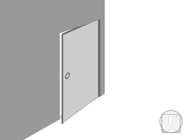
How do you open a stuck drawer (translatory motion joint)?


How do you open a stuck hinged door (rotary motion joint)?
Do we use the same method of opening a drawer to open the door?


How about in a human body joint? 


Have you ever questioned why we glide linearly on our peripheral joint (rotary motion joint)? 


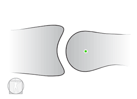
Controversy of Convex-Concave rule
The convex-concave rule, introduced by Kaltenborn into manual therapy, is a didactic simplification of the lever law, during rotatory movements of the joints. However there is much debate in the literature about determining the direction of joint mobilization. The joint axis direction of movement in osteokinematics is different than Kaltenborn’s Convex-Concave rule in determining gliding direction in arthrokinematics.

Joint Surface contact point (Arthrokinematics)
Glide – One point on one joint surface contacts new points on the other joint surface
Roll – New points on each surface come into contact throughout the motion
Spin – The same point on each surface remains in contact with each other
Rotation (New terminology by Dr. Kim) – A single point on each surface contacts multiple points on opposite surface

When the translatoric gliding technique is applied on the convex surface or the concave surface, a single point on the convex surface contacts multiple points on the concave surface. This is completely different on normal rotary motion occurring both on the convex and concave surfaces. Rotary motion occurs when a single point on each surface contacts multiple points on opposite surface.

Angular Joint Mobilization® Concept
Conventional joint mobilization is a translatoric gliding technique performed on a rotary motion joint or on curved translatory motion joint that doesn’t follow osteokinematics and arthrokinematics.

Dr. Kim’s concept of joint mobilization is a rotary technique that has to be applied to a restricted rotary motion joint instead of a translatory gliding technique. During joint mobilization, angular movement (Rotary motion in the joint) should be considered the major movement and translatory movement on the axis or on the articular joint surface should be considered the accessory movement. This insures that the joint structure will be mobilized along the joint surface, stretching other structures effectively and will allow the joint to regain full range of motion more easily since this Angular Joint Mobilization® matches the osteokinematics and arthrokinematics which occur in rotary motion joints.

The bony lever is moving in an angular motion with combined assistive joint mobilization. These include glide, rotation (roll & curved glide), spin, compression, or distraction. This insures that the joint structure will be mobilized along the joint surface, stretching other structures without joint trauma. Joint mobilization has to be multi directional so that the joint structure becomes optimally flexible, enabling it to move in all directions without limitation.

Applying the Angular Joint Mobilization® Technique:
Step 1 - Primary Joint Mobilization - arc of movement utilizing a long lever arm as much as possible to the end range with overpressure to tolerance. This is passive most of the time.
Step 2 - Assistive joint mobilization (Joint Shift/Mobilization) - sustained pressure, or over pressure (glide/rotation/spin/compression/distraction)
Step 3 - Combined movement (Advanced) – Physiological movement to move all direction w/o limitations.

Ex: Knee flexion overpressure - primary joint mobilization
Joint shift (sustained glide/compression/distraction), rotation/spin - assistive joint mobilization
 

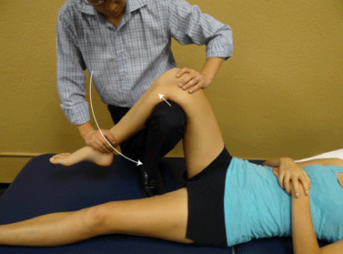
Knee Flexion Angular Joint Mobilization-Anterior shift


Angular Joint Mobilization® Direction (Primary joint mobilization)
It follows the direction of limited lever arm motion (for ex: if shoulder flexion is limited, then the direction of primary joint mobilization is flexion.).

Angular Assistive joint Mobilization Direction
The direction of Angular Assistive joint mobilization can be affected by the joint space, joint surface, capsule, ligaments, muscle (tendon), loss of or defect in the stabilizing mechanism in the joint, pain, muscle spasm, loss of proprioception associated with dysfunction, imbalance/incoordination of the musculature, etc.

The angular assistive joint shift (mobilization) direction doesn’t follow Kaltenborn’s convex concave rule at all.

Helpful tips for determining the angular assistive joint mobilization direction:
• Always apply Primary Joint Mobilization (arc of movement with over pressure) before apply shift and see whether ROM increase or not.
• Apply shift (sustained glide most of the time/compression/distraction) or rotation/spin from mid range (slack) to pathological end range while arc of movement on the lever arm is applied
• Identify the angular assistive joint mobilization direction that provides the most increased ROM
• Identify the most restricted angular assistive joint mobilization direction and provides the most joint structure stretching and increased ROM. (If it is hard to identify this direction, and then simply ask the patient which one produces the most stretching and lesser pain.)
• The joint structure has to be freely moving in all accessory movement directions at every angle of the lever arm.
• Shift in the opposite direction after mobilizing in one direction. (For ex: Glenohumeral joint abduction joint mobilization with posterior shift then abduction mob with anterior shift.)
• Avoid impingement, anatomical limit, or severe pain

The Angular Joint Mobilization® Treatment Plane is not parallel to the concave joint surface because concave joint surface is not flat surface(curved surface) and convex-concave joint is a rotary motion joint not a translatoric motion joint.

Primary Joint Mobilization is applied at the pathological end range of motion, but stops before the anatomical end range of motion. This therapeutic technique should not produce sharp pain, even with over pressure
1. Grade I - In a painful joint, greater rotary oscillations are applied with slight overpressure. Like a swinging pendulum, at the beginning and in the mid part of the arc movement, the joint tissues are on slack. At the end of the arc movement, joint tissue stretching occurs.
2. Grade II - In a non-painful joint, lesser rotary oscillations are needed with more over pressure. At the beginning of the arc movement, the joint tissues are on slack. In the mid (end range of pathological joint) and at the end of the arc movement (over pressure at the end of pathological joint), joint tissue stretching occurs.

Assistive joint shift/mobilization- Sustained pressure / Overpressure:
(Glide /rotation / spin / compression / distraction)
1. Sustained pressure (Shift) – Apply pressure at the end of pathological accessory range to tighten the tissues and sustain
2. Overpressure: Apply overpressure at the end of pathological accessory range to stretch while Primary Joint Mobilization are applied and then come back to the slack position.

Differences between Angular Joint Mobilization® and MWM6 (by Brian Mulligan)

Angular Joint Mobilization® Mobilizations with movement
Can be painful within patient tolerance Pain free
Accessory joint mobilization is not parallel to the concave joint surface Accessory joint mobilization is parallel to the concave joint surface
Can apply any accessory movement (glide/rotation/spin/compression/distraction) – Multidirectional movement Only one direction of accessory joint glide.
Usually passive but can be active with end range overpressure Active or resisted with end range overpressure


Angular Joint Mobilization® indications: All joints with restricted range of motion regardless if capsular or non capsular patterns exist, prosthetic joints (ex: Total knee arthroplasty, etc.).

Angular Joint Mobilization® Contraindications are consistent with the general contraindications for conventional joint mobilizations. These are well described by other authors in various manual therapy books and from the internet.


For more information on Angular Joint Mobilization® please visit www.kiminstitute.org.


Last revised: July 20, 2014
by Younghoon Kim, PT, DPT, OCS, CSCS


REFERENCES
1) Levangie PK, Norkin CC. Joint Structure and Function. A Comprehensive Analysis(ed. 5). F.A. Davis; 2011.
2) Kaltenborn FM. Manual mobilization of the joints. In: The extremities, vol. I. Oslo 7th Edition, Norli Oslo (Norway); 2011.
3) Taber's Cyclopedic Medical Dictionary,2005,p1544
4) Taber's Cyclopedic Medical Dictionary,2009, Issue 21, p192
5) Poppen NK, Walker PS. Normal and abnormal motion of the shoulder. Journal of Bone & Joint surgery Am. 1976;58:195-201.
6) Ludewig PM, Cook TM. Translations of the humerus in persons with shoulder impingement symptoms. Journal of Orthopadic & Sports Physical Therapy. 2002;32:248-259.
7) Matsuki K, et al. Dynamic in vivo glenohumeral kinematics during scapular plane abduction in healthy shoulders. Journal of Orthopadic & Sports Physical Therapy. 2012;42:96-104.
8) Gohlke, FE, et al. Influence of T-shift capsulorrhaphy on rotation and translation of the glenohumeral joint: an experimental study. Journal of Shoulder Elbow Surgery. 1994;3:361-370.
9) Howell SM, et al. Normal and abnormal mechanics of the glenohumeral joint in the horizontal plane. Journal of Bone and Joint Surgery. 1988;70A: 227-232.
10) Soslowsky LJ, et al. Quantitation of in situ contact areas at the glenohumeral joint: a biomechanical study, Journal of Orthopedic Research.1992;10: 524-535.
11) Harryman D, et al. Translation of the humeral head on the glenoid with passive glenohumeral motion. Journal of Bone & Joint surgery Am. 1990;72A: 1334-43.
12) Novotny JE, et al. Normal kinematics of the unconstrained glenohumeral joint under coupled moment loads. Journal of Shoulder and Elbow Surgery. 1998;7:629-639.
13) Teyhen DS, et al. Rotator cuff fatigue and glenohumeral kinematics participants without shoulder dysfunction. Journal of Athletic Training. 2008;43:352-358.
14) Graichen H, et al. Effect of abducting and adducting muscle activity on glenohumeral translation, scapular kinematics and subacromial space width in vivo. Journal of Biomechanics. 2005;38:755-760.
15) Chen SK, et al. Radiographic evaluation of glenohumeral kinematics: A muscle fatigue model. Journal of Shoulder and Elbow Surgery. 1999;8:49-52.
16) Deutsch A, et al. Radiologic measurement of superior displacement of the humeral head in the impingement syndrome. Journal of Shoulder and Elbow Surgery. 1996;5:186-193.
17) Hallström E, et al. Shoulder kinematics in 25 patients with impingement and 12 controls. Clinical Orthopaedics and Related Research. 2006;448:22-27.
18) Nishinaka N, et al. Degermination of in vivo glenohumeral translation using fluoroscopy and shape-matching techniques. Journal of Shoulder and Elbow Surgery. 2008;17:319-322.
19) Bey MJ, et al. Measuring dynamic in-vivo glenohumeral joint kinematics: Technique and preliminary results. Journal of Biomechanics. 2008;41:711-714
20) Baeyens J-P, van Roy P, Clarys JP. Intra-articular kinematics of the normal glenohumeral joint in the late preparatory phase of throwing: Kaltenborn’s rule revisited. Ergonomics 2000;43(10):1726–37.
21) Baeyens J-P, van Roy P, de Schepper A, Declercq G, Clarijs J-P. Glenohumeral joint kinematics related to minor anterior instability of the shoulder at the end of the late preparatory phase of throwing. Clinical Biomechanics 2001;16:752–7.
22) Cattrysse E, Baeyens J-P, van Roy P, van de Wiele O, Roosens T, Clarys J-P. Intraarticular kinematics of the upper limb joints: a six degrees of freedom study of coupled motions. Ergonomics 2005;48(11–14):1657–71.
23) Baeyens J-P, van Glabbeek F, Goossens M, Gielen J, van Roy P, Clarys J-P. In vivo 3D arthrokinematics of the proximal and distal radioulnar joints during active pronation and supination. Clinical Biomechanics 2006;21:S9–12.
24) Brandt C, Sole G, Krause MW, Nel M. An evidence-based review on the validity of the Kaltenborn rule as applied to the glenohumeral joint. Man Ther. 2007 Feb;12(1):3-11. Epub 2006 Jun 13.
25) Schomacher J. Response to: Brandt C, Sole G, Krause MW, Nel M. An evidencebased review on the validity of the Kaltenborn rule as applied to the glenohumeral joint. Manual Therapy 2007;12(1):3–11. Manual Therapy 2008;13(1):e1–2.
26) Johnson AJ, Godges JJ, Zimmerman GJ, Ounanian LL. The effect of anterior versus posterior glide joint mobilization on external rotation range of motion in patients with shoulder adhesive capsulitis. J Orthop Sports Phys Ther. 2007 Mar; 37(3):88-99.
27) Schomacher J. The convex-concave rule and the lever law. Man Ther. 2009 Oct; 14(5):579-82. Epub 2009 Mar 5.
28) Mulligan, BR; Manual Therapy 'NAGS', 'SNAGS', 'MWMS' etc.(6th Ed), Orthopedic Physical Therapy Products, 2010


Terms & Conditions

Please review our terms and conditions carefully before utilization of the Site. The information on this Site is for informational purposes only and should in no way replace a conventional visit to an actual live physical therapist or other healthcare professional. It is recommended that you seek professional and medical advise from your physical therapist or physician prior to any form of self treatment.


Advertisement
.

Advertisement

.