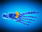
|
Conditions and Treatments - Description and Management of Accessory Navicular Syndrome ׀ by Bill Lyon, PT, DPT, CSCS |

Bill Lyon, PT, DPT, CSCS, USAW-L1 received his doctor of physical therapy degree from the University of Wisconsin - Milwaukee. He has more than 9 years of experience in performance training and strength & conditioning and is a Certified Strength and Conditioning Specialist through the NSCA as well as a Level one Olympic lifting coach through United States Weightlifting. Bill is a physical therapist with United Hospital System in Kenosha where he works primarily in an outpatient physical therapy setting.
|
|
Description and Management of Accessory Navicular Syndrome |
| . |
 The accessory navicular is an accessory bone that can
develop and cause enlargement of the plantar medial
navicular. Generally there are three presentations of
the accessory navicular; as a sesmoid bone in the
posterior tibialis tendon, articulated with the
navicular, or fused to the navicular. An accessory
navicular is a common accessory formation of the foot
and is present in ~ 10% of the population. It is more
common in females vs males and is commonly bilateral
(70% of cases). Accessory naviculars may often be benign
and cause no pain or issue, but when pain and
dysfunction do occur, it is referred to as accessory
navicular syndrome. The accessory navicular is an accessory bone that can
develop and cause enlargement of the plantar medial
navicular. Generally there are three presentations of
the accessory navicular; as a sesmoid bone in the
posterior tibialis tendon, articulated with the
navicular, or fused to the navicular. An accessory
navicular is a common accessory formation of the foot
and is present in ~ 10% of the population. It is more
common in females vs males and is commonly bilateral
(70% of cases). Accessory naviculars may often be benign
and cause no pain or issue, but when pain and
dysfunction do occur, it is referred to as accessory
navicular syndrome. In accessory navicular syndrome, there is dysfunction that is caused by the displacement of the normal posterior tibialis tendon insertion. With the displaced tendon attached to the accessory bone, there is a loss in support of the medial arch of the foot. This may lead to pes planus, although a cause and effect relationship between accessory navicular and pes planus has not been proven. Presence of articulating or fused accessory naviculars can also lead to posterior tibialis tendonopathy due to the change in insertion and increased stress to the tendon. This condition is most common in young (10-30 year old) females with complaints of pain in the midfoot, arch, and or directly over the area of navicular prominence. The prominence will likely be tender to palpation and enlarged. Common complaints are pain with tighter footwear and pain with prolonged standing or walking. Often there will also be pes planus and pain over the posterior tibialis tendon as well. A lateral oblique view radiograph may assist with diagnosis, although the accessory may not be distinct if ossified to the navicular. Differential diagnoses to consider would be stress fracture, tendonitis, and Kohler’s disease. Non-surgical treatment often includes strengthening of the intrinsic and extrinsic muscles of the foot and ankle, primarily those that assist in lifting the arch, strengthening of the external rotators of the hip to assist with foot mechanics, modalities for pain relief and decreasing inflammation, and orthotic management to decrease the stress to the posterior tibialis tendon and unload the pressure to the navicular its self. Often anti-inflammatory medications are used in oral and injection forms. Some cases may be immobilized for a time period in CAM walker boot to decrease stress during weight bearing. In cases not responding to conservative therapy, surgical approaches may be used. A simple excision of the accessory bone prominence relieves symptoms for many patients. A second option is a kidner procedure, which involves excision of the prominence, as well as advancement of the posterior tibialis tendon.
Last revised: December 28, 2016
References |
|
|
|
|


